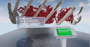
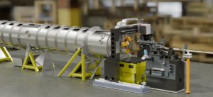
Author: Lund University

Segmenting Glomeruli and other structures from synchrotron X-ray microtomography datasets

Segmenting Glomeruli and other structures from synchrotron X-ray microtomography datasets

Diabetes is a long-lasting health condition with serious complications. We studied a new group of rats with a specific genetic mutation linked to diabetes. Regardless of their gender and blood sugar levels, these rats were overweight and had larger livers, hearts, and kidneys compared to normal rats. A study of the size of Bowmans space (difference between outer perimeter and Tuft of their glomeruli) can reveal tendencies toward having or developing cancer. In that respect advanced machine learning approaches are used to automatically segment and evaluate 3D volumetric information of many of the structures found within the tomography scans collected.

Butterflies in Virtual Reality: developing workflows for efficient morphological segmentation and analysis of X-ray microtomography Datasets

Butterflies in Virtual Reality: developing workflows for efficient morphological segmentation and analysis of X-ray microtomography Datasets

Initially an InfraVis pilot project, the VR lab at LTH has worked with Sridhar Halali, a researcher at the LU Dept. of Biology. As part of his work to understand how butterflies have evolved in response to environmental changes, Dr Halali has collected a dataset of microCT scans of butterflies. The specific trait of interest is in this case the shape of the head. It can, however, be a greatly time-consuming task to isolate body segments in a large 3-dimensional dataset using the 2D interface of the standard software (3D Slicer). This is where the VR Lab comes in, analyzing the problem, building on existing plugins for visualization and interaction in VR, and parallel processing to implement a solution to facilitate the task.