3D mapping of cortical changes leading to epileptic seizures
InfraVis User
My Andersson
InfraVis Application Expert
Jonas Ahlstedt
InfraVis Node Coordinator
Anders Sjöström, Emanuel Larsson
About
Epilepsies are a family of devastating disorders characterized by spontaneous, recurrent seizures. It is one of the most common brain disorders and considered the most burdensome neurologic disorder worldwide. Available pharmacological treatments are only symptomatic, often with side effects and fail to adequately control seizures in one third of patients.
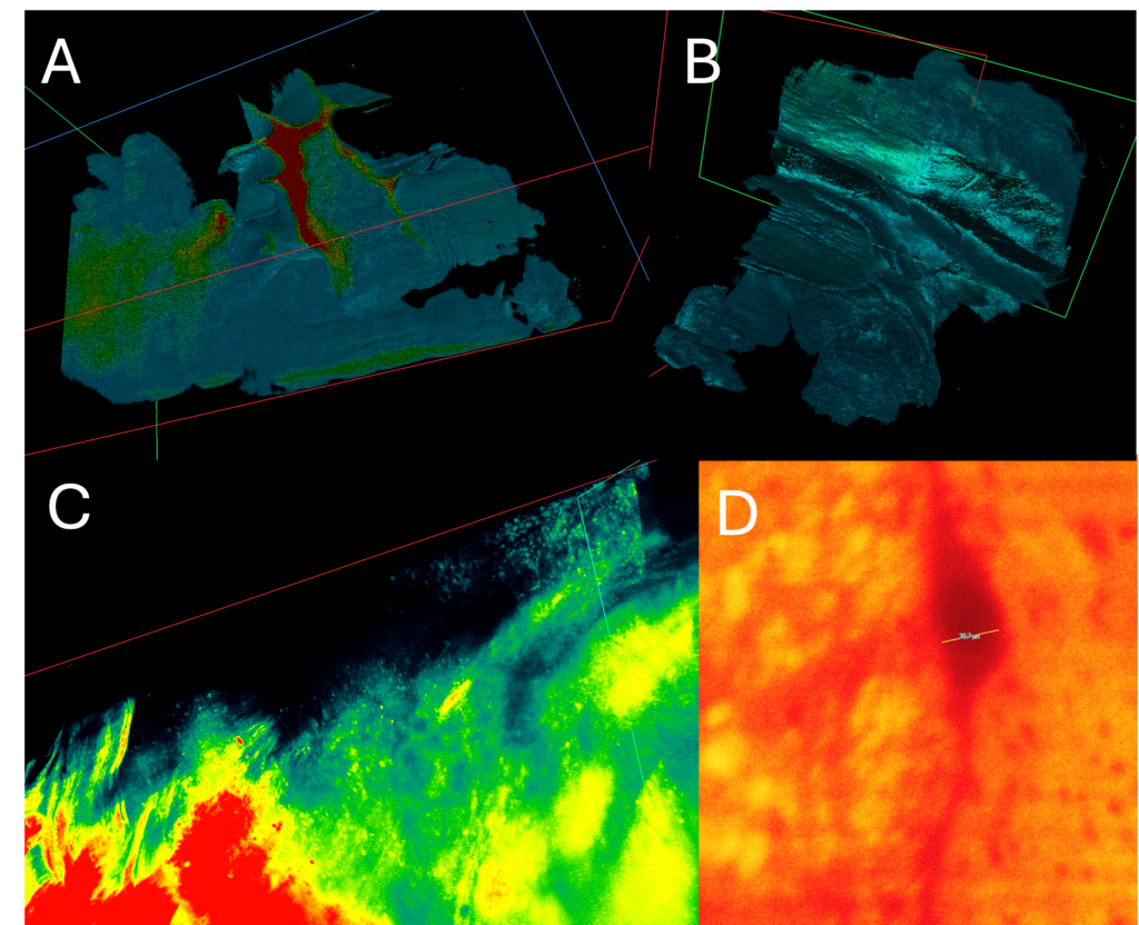
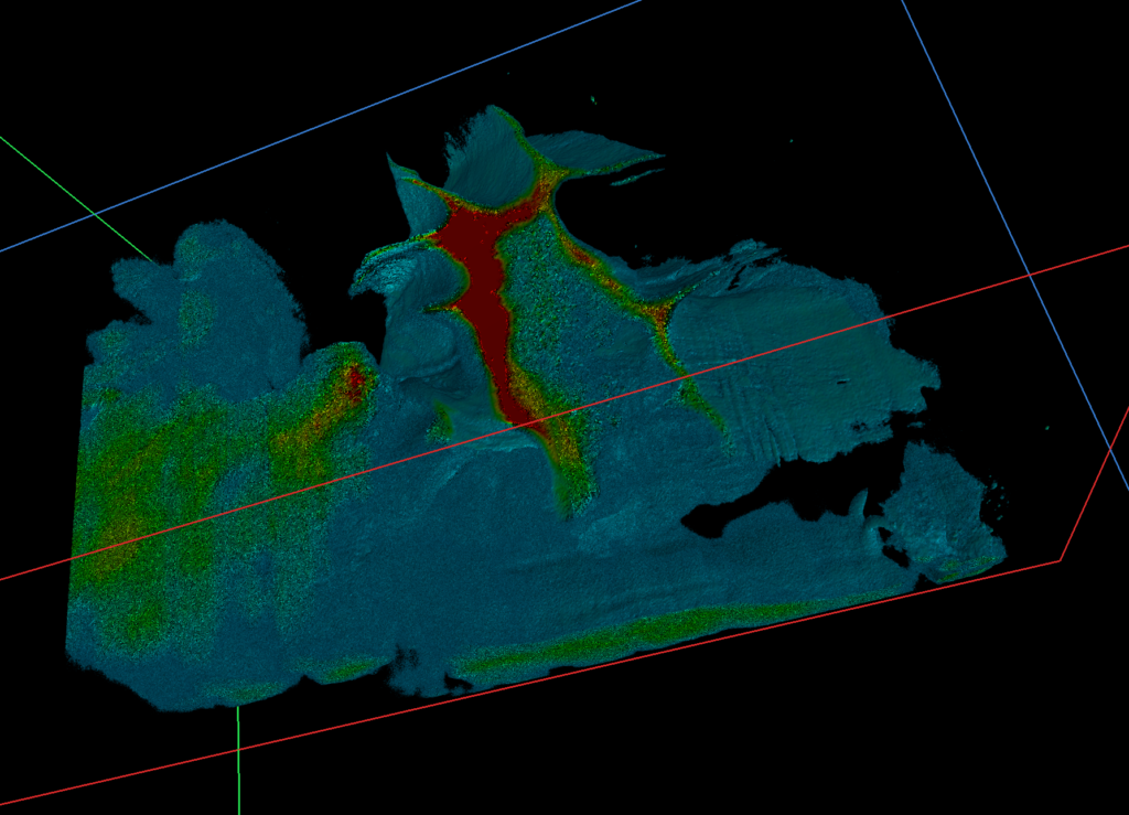
A 3D-map of the structural changes, underlying the pathological hyperexcitable network leading to seizures, could give us the directions needed to reverse and thus cure epilepsy.
Lightsheet data is notorious for its capacity to provide high-resolution image stacks and can therefore prove a challenge in rendering the 2D information as a 3D volume. In this case each plane in the stack consisted of stitched images making each stack up to 500 GB in size. The only software seemingly capable of converting the stacks and displaying them comfortably (or at all) with real-time manipulation was ArivisVision4D. A custom MATLAB script was developed to convert the images from JPEG2000 (lossless format) to TIFF stacks and the conversion to 3D was performed using ArivisVision4D.